


Wound closure techniques have advanced to include synthetic sutures, staples, tapes, and adhesives.
Synthetic sutures and standardized materials enhance aesthetic results in engineering.
Topical adhesives, surgical staples, and tapes supplement sutures in wound closure techniques.
Proper materials and technique ensure optimal healing. Four wound healing phases studied at cellular and molecular levels.
The four phases are: Hemostasis, Inflammation, Proliferation, Maturation phase
Initial injury disrupts blood vessels and exposed collagen triggers hemostasis with normal thrombotic function.
Epithelial cell migration occurs in 12-24 hours, with new tissue formation over 10-14 days.
Wound Irrigation
Debridement Tools
Clamps
Suturing and Closure
Dressings
Tape or Bandages
Adhesives
Patient Preparation:
Inform the patient about surgery risks, benefits, recovery, pain management, restrictions, and physical therapy.
Ensure understanding before obtaining written informed consent.
A mixture of one part sodium bicarbonate and ten parts local anesthesia with epinephrine can reduce burning during lidocaine infiltration.
Patient Positioning:
Position patient for best wound access and visibility.
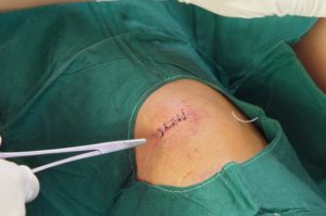
Figure 1. Wound suture
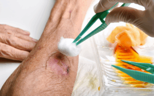
Figure 2. Wound dressing
Closure by secondary intention effectively addresses wounds on concave head and neck areas particularly those at infection risk.
Results are aesthetic and functional to avoid complex flap or skin graft procedures.
The final scar is less visible in older, lighter-skinned patients, and suitable with other techniques.
Deep sutures eliminate dead space, relieve tension, ensure wound edge alignment, and contribute to final eversion of the wound.
For head and neck wound closure, use 5-0 or 6-0 nonabsorbable Prolene, nylon, or absorbable catgut sutures carefully.
Eversion of skin edges prevents scar depression and place simple suture knots away from wound edges.
Running Locking Suture: Continuous suture with loops locked for added strength.
Vertical Mattress Sutures: It provides strong eversion of edges to prevent depressed scars.
Horizontal Mattress Sutures: It distributes tension over a wider area.
Approximate wound edges to initiate healing, avoid tight sutures that compromise blood flow to cause tissue necrosis, scarring, and unsatisfactory cosmetic outcomes.
Sutures tied tightly today will become tighter tomorrow. Despite careful tissue handling to cause swelling and increased tension at the suture site.
Subcuticular suture procedure:
Sutures can be intradermal either simple or running with the needle placed horizontally in the dermis without skin penetration.
The buried knot in a simple suture minimizes wound tension, while continuous subcuticular stitches allow suture ends to be taped without knots.
Complications:
Wound dehiscence
Chronic wound formation
Hematoma and seroma formation
Suture-related issues
Necrosis
Scarring
LEARNING & CME
View All
Advanced
Cardiovascular
Life Support

Basic Life
Support

Pediatric
Advanced Life
Support

Neonatal
Resuscitation
Program

Annual Stroke
Center
Continuing
Education

Opioid and Pain
Management

National
Institutes of
Health Stroke
Scale

Basics of
Electrocardiography