


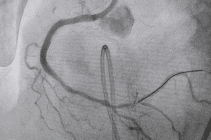
Selective arterial embolization is a minimally invasive medical technique that selectively blocks blood vessels in the arterial system. It involves in injecting embolic materials into the system blocking blood flow to targeted tissues or organs. The process typically involves a catheter threaded through blood vessels with imaging methods like fluoroscopy used to guide the delivery of embolic agents.
The angiography suite includes a fluoroscopy machine, digital subtraction angiography system, monitor, catheters, embolic agents, vascular access devices, imaging contrast agents, fluoroscopy table, anesthesia and monitoring equipment, sterile drapes and gowns, syringes and contrast injectors, and post-procedural imaging equipment.
The fluoroscopy machine provides real-time X-ray images while the digital subtraction angiography system enhances blood vessel visibility.
Catheters are used for navigating and visualizing blood vessels while embolic agents block blood vessels and control bleeding.
Vascular access devices include intubator sheaths, needles, and guide catheters.
Anesthesia and monitoring equipment monitor vital signs, sterile drapes and gowns to ensure a sterile environment during the procedure.
Post-procedural imaging equipment assesses the effectiveness of embolization.
A comprehensive medical evaluation is conducted that includes a review of past medical history, allergies, and current prescriptions.
Patients should inform their healthcare team about any existing medical conditions especially kidney problems or allergies to contrast dye.
Blood tests may be performed to determine general health status and detect infections.
The provider may adjust or temporarily stop certain medications especially those affecting blood clotting.
Patients should inform their healthcare team about any allergies especially to iodine-based contrast agents.
A consent form is signed before the procedure and outlining the procedure details, risks, and benefits.
Patients should dress comfortably and loosely on the day of the procedure by the team instructions.
Step 1:
Seldinger procedure which involves placing a catheter via the common femoral artery which was utilized for digital subtraction angiography and selective arterial embolization under local anesthesia.
Selective arterial embolization
Step 2:
Glubran 2 surgical adhesive was mixed with lipiodol ultra fluid at a 33% dilution and is administered with a 5% glucosate solution to create a sandwiched injection by preventing polymerization in blood contact.
Step 3:
The process involved is three phases starting with Digital Subtraction Angiography to create a vascular map of the PT anatomical region.
Step 4:
The second phase involved selective catheterizations using a coaxial catheter system which is followed by embolization of lesion-supplying vessels by Digital Subtraction Angiography to confirm the occlusion effectiveness and assess any remaining pathological blood flow.
Step 5:
After the procedure was completed a post-procedure PT and Digital Subtraction Angiography was performed to evaluate the occlusion efficacy.
Pre-procedural Tests:
Blood tests are essential for evaluating a patient baseline condition and confirming their ability to withstand surgery.
Angiogram is a crucial imaging technology which is used in embolization that requiring contrast dye injection to view the target area and blood vessels. Post-procedural tests include follow-up imaging to assess the success of the embolization and monitor blood flow in the treated area.
Periodic blood tests are also conducted to monitor any changes in blood parameters especially if the embolization was done to address a specific medical condition.
Selective arterial embolization can lead to ischemia, where blood flow is blocked to a specific area that potentially causing tissue damage or organ dysfunction and the severity of which depends on the target area that interrupts the duration.
Invasive surgery particularly embolization can introduce foreign materials that potentially leading to infection. Although the risk is generally low and it is crucial in immunocompromised individuals for their safety.
Rare cases of allergic reactions may occur with embolic agents and radiopaque contrast agents used in imaging procedures.
Accidental embolization or damage to nearby structures can be dangerous but necessitating careful planning and imaging guidance will minimize this risk.
During embolization there is a risk of perforating or rupturing blood vessels particularly if the vessels are fragile or diseased.
LEARNING & CME
View All
Advanced
Cardiovascular
Life Support

Basic Life
Support

Pediatric
Advanced Life
Support

Neonatal
Resuscitation
Program

Annual Stroke
Center
Continuing
Education

Opioid and Pain
Management

National
Institutes of
Health Stroke
Scale

Basics of
Electrocardiography