


Renal imaging may be defined as the process of localizing and visualizing the kidney and all the structures that may be attached to it. This is useful when deciding on the course of action in relation to several renal disorders such as obstructive uropathy, tumors, infections and congenital abnormalities.
Ultrasound (US): Involves the production of images of the kidneys with the help of high-frequency sound waves. This is a non-destructive and commonly employed method.
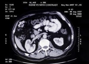
Computed Tomography (CT) Scan: Appears to utilize X-ray and computers to take a picture of the kidneys and tissues in another plane. Offers clear images and can be used in diagnosing kidney stones, tumors, and constituents of the urinary system obstructs. CT urography is a specific type of CT scan that focuses particularly on the system of the urinary tract.
Magnetic Resonance Imaging (MRI): Creates clear pictures of the kidneys using strong magnetic fields and radio waves that do not harm the patient. Produces image with clearer resolution and does not use radiation for reviewing various conditions of the kidneys and to differentiate between various types of tissue.
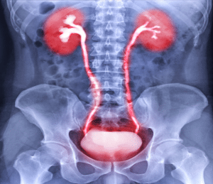
Intravenous Pyelogram (IVP): An older technique in this system where a contrast dye is administered intravenously, and fluoroscopic images are taken while the dye goes through the renal and urinary system. Contains details of the kidney and the urinary system and how they work and their formation.
Renal Scintigraphy (Nuclear Medicine): It involves the use of radioactive tracers to determine the efficiency of kidneys as well as its build or formation.
Vital Signs Monitor: To measure the patient’s pulse rate, blood pressure, SpO2 and respiratory rate at a frequency that will enhance patient care.
Intravenous (IV) Access: Intravenous catheters for the delivery of contrast media, medications as well as fluids.
Emergency Resuscitation Equipment: Staples which are defibrillator, oxygen, and emergency drugs (epinephrine, antihistamines for allergic reactions and other complications).
Contrast Media Injectors: Pump-like for injecting contrast media into the body for CT, MRI, and angiography.
Ultrasound
Ultrasound Machine: Delivered with numerous openly/separately connectable probes (transducers) for multiple imaging purposes.
Gel: Conductive gel that is smeared on the skin surface to enhance a good contact between the probe and the skin.
Patient Positioning Equipment: Pillows, wedges, straps to help maintain the patients comfort for imaging as well as positioning him/her in the best possible manner.
Computed Tomography (CT) Scan
CT Scanner: An apparatus that employs rays to form clear sectional pictures.
Contrast Media: Iodated more effective for improving image quality of the scans.
Lead Shields: Attire and equipment that are worn by workers to protect other parts of the body from radiation.
Radiation Dosimeters: Equipments used in measurement of the amount of radiation dosage in any environment.
Magnetic Resonance Imaging (MRI)
MRI Scanner: An apparatus that employs an electromagnetic field and radio oscillations to generate clear pictures.
Gadolinium-based Contrast Agents: For improving the quality of the MRI such as the image contrast.
Ear Protection: Headphones or ear plugs to shield yourself from the sound produced by MRI equipments.
Positioning Cushions: Because of the patient comfort and positioning of the patient in the correct position during the scan.
MRI Scan
Intravenous Pyelogram (IVP)
X-ray Machine: These are utilized in imaging the urinary tract with an objective of observing lining of the opening of the ureter into the bladder.
Iodinated Contrast Media: Given intra-venously for the purpose of outlining the kidneys, the ureters and the bladder.
Compression Devices: Applied prior to the procedure to decrease the rate of urination and to improve the visibility of the urinary tract.
Ultrasound
The patient may be asked to drink water to ensure a full bladder, which can improve imaging of the kidneys. The patient lies on an examination table, usually in a supine position, but sometimes prone or on their side. Conductive gel is applied to the patient’s skin over the area to be examined. The ultrasound transducer (probe) is moved over the skin to capture real-time images. Different transducers may be used for various depths and resolutions.
Computed Tomography (CT) Scan
Fasting for a few hours before the scan. Hydration may be encouraged to help protect the kidneys. The patient lies on a motorized table that slides into the CT scanner. Intravenous contrast may be given to enhance image quality. Oral contrast may be used in some cases. The scanner rotates around the patient, taking multiple X-ray images that are then reconstructed into cross-sectional slices.
Magnetic Resonance Imaging (MRI)
Screening for contraindications (e.g., metallic implants). Fasting may be required if contrast is used. The patient lies on a table that slides into the MRI machine. Gadolinium-based contrast may be injected intravenously. Magnetic fields and radio waves are used to create detailed images. The patient must remain still, and the procedure can take longer than a CT scan.
Intravenous Pyelogram (IVP)
Fasting and bowel preparation may be required. Hydration is encouraged. The patient lies on an X-ray table. Iodinated contrast is injected intravenously. X-ray images are taken at various time intervals to capture the excretion of the contrast through the kidneys, ureters, and bladder.
Approach considerations
Ultrasound (US):
Advantages: Can be performed without a need to use contrast agents, does not use ionizing radiation; is valuable in the initial evaluation of the renal pathology; it also assesses the renal size, shape, and architecture and may detect cysts, hydronephrosis, and stones.
Limitations: Lacks ability to penetrate deeply into the body and visualizing small lesions; less clear as compared to other techniques.
Computed Tomography (CT):
Advantages: Produces good spatial and contrast detail; helpful for attempting to assess the presence of renal abnormalities and urinary tract obstruction of over 5mm in size as well as tumor and stones assessment and renal artery patency.
Limitations: It uses ionizing radiation, and the contrast agents could be contraindicated in patients who have renal problems.
Magnetic Resonance Imaging (MRI):
Advantages: No ionizing radiation, particularly useful for soft tissues; good for distinction of renal masses, congenital malformations and vascular lesion.
Limitations: More expensive and may be contraindicated in certain types of implant patients or patients who have claustrophobia; contrast agents may also be hazardous for patients with severe renal disease.
Renal Scintigraphy (Nuclear Medicine):
Advantages: It helps evaluate the renal functionality and blood flow and can be used to determine if the renal problems are obstructive or non-obstructive.
Limitations: Assists in blood vessel imaging, provides a lower detail resolution compared to CT or MRI scans.
Serum Creatinine: Tests the concentration of creatinine in the blood – metabolism byproduct of muscles. Hyperkalemia indicates the possible renal insufficiency.
Blood Urea Nitrogen (BUN): Tests kidney function by checking of the level of urea nitrogen in the blood. Marked/abnormal elevation might be suggestive of renal impairment.
Urinalysis: Screens for infection, blood, protein, glucose or any other substances that could point to problems with the kidney.
Urine Albumin-to-Creatinine Ratio: An index that evaluates the level of albumin (a type of protein) in urine with the level of creatinine in the blood to identify the kidney disease.
Electrolytes (e. g., Sodium, Potassium, Calcium): Abnormalities can indicate kidney disorders as kidneys help in the balance of these chemicals.
Glomerular Filtration Rate (GFR): A simple formula that predicts the level of kidney function with the help of serum creatinine, age, gender, and race factors. This shows level of how effective the kidneys are in filtering the blood.
Ultrasound
No significant complications: Ultrasound has been rated as safe for use and has few side effects. Some people can develop some level of discomfort from the probe or the gel that is used, but these are usually not very serious.
CT scan
Contrast-related reactions: Contrast agents when used may cause allergic reactions which start from mild to severe. It is also useful to mention that the procedure can cause contrast-induced nephropathy in patients with kidney disease.
Radiation exposure: In performing CT Scans, ionizing radiations are used and these when taken repeatedly may lead to high risks of cancer.
MRI
Contrast-related reactions: Like CT, the intrusive substances used for MRI, such as gadolinium, also have same allergic reaction or nephrogenic systemic fibrosis in the patients of chronic renal disease.
Metallic implants: In MRI examination some patients with metallic implant and devices develop complications due to the fact that MRI uses strong magnetic fields.
LEARNING & CME
View All
Advanced
Cardiovascular
Life Support

Basic Life
Support

Pediatric
Advanced Life
Support

Neonatal
Resuscitation
Program

Annual Stroke
Center
Continuing
Education

Opioid and Pain
Management

National
Institutes of
Health Stroke
Scale

Basics of
Electrocardiography