


Intravascular ultrasonography (IVUS) is a medical imaging technique which involves the use of high-frequency sound waves (ultrasound) to visualize the interior of blood vessels and assess the structure and condition of the vascular walls. It provides detailed, real-time images of blood vessels, helping healthcare providers diagnose and treat various cardiovascular conditions.
IVUS was first introduced in the late 1980s and has since undergone significant advancements in technology and clinical applications. The initial IVUS systems were limited in their imaging quality and required invasive procedures, but modern IVUS technology has become more user-friendly, safe, and informative.
IVUS uses a small ultrasound transducer mounted on the tip of a catheter, which is inserted into a blood vessel, typically an artery or a vein. Transducer emits high-frequency sound waves that bounces off the surrounding tissues and structures within the vessel. The reflected sound waves are captured and processed to create cross-sectional images of the vessel and its walls.
IVUS is commonly used in interventional cardiology to assess coronary arteries during angioplasty and stent placement procedures. It helps cardiologists evaluate the extent of arterial blockages and the appropriateness of stent placement. It is also used in peripheral vascular procedures to assess vessels in the legs and arms, particularly in cases of peripheral artery disease (PAD). IVUS is valuable for evaluating the vascular anatomy and condition in organ transplant surgery.
- Patient Assessment: Conduct a comprehensive assessment of the patient’s medical history, current condition, and indications for IVUS. Ensure that the patient’s clinical presentation justifies the procedure.
- Informed Consent: Obtain the informed consent form from patient, providing a clear explanation of the procedure, its purpose, potential risks, and benefits.
- Allergies and Medical History: Assess the patient for allergies, especially to iodine-based contrast agents, as well as any underlying medical conditions that may influence the choice of contrast agent or affect the procedure’s safety.
- Renal Function: Evaluate the patient’s renal function, as some contrast agents used in IVUS contain iodine, and impaired renal function can affect the choice of contrast agent and the risk of contrast-induced nephropathy.
- Hemodynamic Stability: Ensure that the patient is hemodynamically stable before the procedure. Unstable patients may need stabilization before IVUS.
- Coagulation Profile: Assess the patient’s coagulation profile, especially in cases where anticoagulants or antiplatelet agents are involved, to mitigate the risk of bleeding complications.
- IVUS Catheter: Select the appropriate IVUS catheter based on the vessel of interest, the procedure’s purpose, and the desired imaging depth. Catheters may vary in size and frequency (e.g., 20 MHz, 40 MHz).
- IVUS Console: Ensure the availability and proper functioning of the IVUS console, which includes the ultrasound transducer, imaging software, and controls.
- Contrast Agent: Prepare the appropriate contrast agent, which is used to enhance vascular visualization during the procedure. Ensure that the patient is not allergic to the chosen contrast agent.
- Guidewires and Catheters: Have the necessary guidewires and guiding catheters available for vascular access and navigation.
- Hemostatic Equipment: Prepare hemostatic equipment for vessel closure and minimizing bleeding risk post-procedure.
- Monitoring Equipment: Equip the procedure room with standard monitoring devices, including electrocardiogram (ECG), blood pressure, and oxygen saturation monitors.
- Sterile Drapes and Gowns: Ensure that sterile drapes and gowns are available to maintain aseptic conditions during the procedure.
- Personal Protective Equipment: Healthcare providers should wear appropriate personal protective equipment, including sterile gloves and gowns, masks, and eye protection.
- Local Anesthesia and Sedation: Prepare local anesthetic agents for pain management and any necessary sedation or anesthesia, based on the patient’s comfort and the complexity of the procedure.
Step:1 Approach Considerations:
- The specific approach for Intravascular Ultrasonography (IVUS) depends on the vessel of interest and the clinical indication. Accessing the vessel may involve femoral, radial, or brachial artery puncture.
- Pre-procedural evaluation includes patient assessment, informed consent, allergy assessment, and assessment of coagulation profile and renal function.
Step:2 Normal Arterial Appearance:
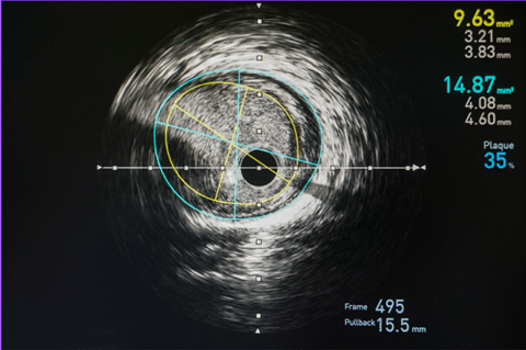
- IVUS imaging provides cross-sectional images of the arterial wall layers. A healthy artery shows three layers: the intima, media, and adventitia.
- The intima is the innermost layer, followed by the media, composed of smooth muscle cells, and the adventitia, which surrounds the vessel.
Step: 3 Quantitative Measurements:
- IVUS provides precise measurements of vessel size, plaque burden, lumen diameter, and wall thickness.
- Quantitative parameters include lumen area, vessel area, plaque area, minimal lumen diameter, and external elastic membrane (EEM) area.
Step:4 Qualitative Assessment:
- IVUS allows assessment of plaque morphology, such as calcifications, fibrous tissue, lipid pools, and thrombus.
- Plaque characterization helps determine lesion severity and guides treatment decisions.
Step:5 In-Stent Restenosis and Drug-Eluting Stent Implantation:
- IVUS is particularly useful in evaluating stent deployment and apposition. It helps in assessing stent expansion, symmetry, and malapposition.
- In cases of in-stent restenosis, IVUS aids in characterizing the restenotic tissue and guides decisions on further intervention.
- Atherosclerosis: IVUS provides insights into the extent and composition of atherosclerotic plaques. It helps assess vulnerable plaques prone to rupture.
- Coronary Artery Disease (CAD): IVUS is commonly used during PCI to assess the severity of lesions, guiding stent selection, optimizing stent deployment, and evaluating procedural success.
- Peripheral Artery Disease (PAD): IVUS helps in evaluating the severity of arterial stenosis in peripheral vessels, guiding treatment decisions for endovascular interventions.
- Intracranial Aneurysms: While IVUS has been explored in intracranial vessels, its use in assessing intracranial aneurysms is limited due to potential vessel damage risk.
- Renal Artery Stenosis: IVUS can assist in evaluating the degree of renal artery stenosis and guiding decisions for interventions like renal artery stenting.
- Transplant Vessels: IVUS aids in evaluating vessel patency, identifying anastomotic problems, and optimizing vascular anastomosis during organ transplantation.
LEARNING & CME
View All
Advanced
Cardiovascular
Life Support

Basic Life
Support

Pediatric
Advanced Life
Support

Neonatal
Resuscitation
Program

Annual Stroke
Center
Continuing
Education

Opioid and Pain
Management

National
Institutes of
Health Stroke
Scale

Basics of
Electrocardiography