


Hernia reduction involves pushing protruding tissue back into normal position during treatment.
Hernia happens when organ or tissue pushes through weak muscle and connective spot
Hernia repair is a common surgery type. It frequently occurs in abdomen, groin, and near incisions.
Hernias are divided into two main groups as:
Groin hernias
Abdominal hernias
These are further sub-divided as:
Inguinal Hernia: In the groin area
Femoral Hernia: Located just below the inguinal ligament
Umbilical Hernia: It involves part of the intestine
Spigelian hernia: Located at the lateral edge of the rectus abdominis
Manual reduction of a hernia before surgery relieves symptoms and prevent complications. Patient should relax their abdominal muscles, while physician applies pressure to encourage tissue/organ to return properly.
This process may relieve symptoms temporarily but is not a permanent solution.
Cold compress
Scalpel & Forceps
Needle Holders
Surgical Sutures
Anesthesia Equipment
Wound Closure and Dressing
Laparoscopic Instruments
Laparoscope
Trocar and Cannulas
Laparoscopic Suturing Devices
For hernia present in the intestine, bowel preparation may be required to reduce the risk of infection during surgery.
Patients are instructed to fast for 6 to 8 hours prior to surgery.
Patients should understand procedure, benefits, risks, and alternatives for consent.
Place the patient in a Trendelenburg position. (i.e., head down & legs up)
In cases of an inguinal hernia, the patient should be in a supine position.
Step 1: Relax abdominal muscles:
Motivate patients to relax their abdominal wall with gentle pressure and ask them to breathe deep.
Step 2: Application of Gentle Pressure
Slow and steadily apply pressure with your fingers or the palm of hand over the herniated bulge. This should gently guide the contents back through the defect in the abdominal wall.
A. Open Hernia Repair:
Step 1: Anesthesia
General/local anesthesia is administered.
Step 2: Incision
Surgeons will make incisions directly at the site of the hernia.
Step 3: Reduction of Hernia
The surgeon carefully identifies the herniated tissue and gently reduces it back into the abdominal cavity.
Step 4: Hernia Repair
For small hernia cases, the defect may be closed with sutures alone by stitching the edges of the healthy tissue together.
For larger hernia cases, a synthetic mesh is placed over or under the defect to reinforce the weakened area.
Step 5: Closure
The surgical wound is closed in layers. The deeper muscle and fascia are sutured first.
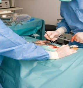
Open Hernia Repair
B. Laparoscopic Hernia Repair:
Step 1: Anesthesia
General anesthesia is administered.
Step 2: Incision and Access
Small incisions are made in the abdominal wall to insert the laparoscope.
The abdomen is inflated with CO2 gas to create space for visualization and manipulation of the herniated tissue.
Step 3: Reduction of Hernia
The herniated contents are observed with the laparoscope and then surgeon gently manipulates the herniated tissue back into the abdominal cavity.
Step 4: Placement of mesh
Synthetic mesh is placed over the hernia and fixed in place with sutures.
Step 5: Closure
The small incisions are closed with sutures.
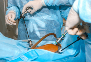
Laparoscopic Hernia Repair
Complications:
Complications of Manual Hernia Reduction are:
Incomplete Reduction
Injury to Herniated Contents
Missed Strangulation
Hematoma or Seroma Formation
Complications of Surgical Hernia Reduction are:
Infection
Bleeding or Hematoma
Seroma
Hernia Recurrence
Bowel Injury or Perforation
Bowel Obstruction
Ischemia or Necrosis of Herniated Tissue
LEARNING & CME
View All
Advanced
Cardiovascular
Life Support

Basic Life
Support

Pediatric
Advanced Life
Support

Neonatal
Resuscitation
Program

Annual Stroke
Center
Continuing
Education

Opioid and Pain
Management

National
Institutes of
Health Stroke
Scale

Basics of
Electrocardiography