


Fallopian tube reconstruction is a surgical procedure which is done with the aim of reconstructing the tubes or prolong the function of the fallopian tubes because these tubes play an important role in fertility as they are the conduits of the eggs from the ovaries to Uterus. This reconstruction is most often advised to women who have tubal blockage due to reasons such as infection, endometriosis, or previous surgeries.
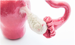
Fallopian tube reconstruction is a surgical procedure which is done with the aim of reconstructing the tubes or prolong the function of the fallopian tubes because these tubes play an important role in fertility as they are the conduits of the eggs from the ovaries to Uterus. This reconstruction is most often advised to women who have tubal blockage due to reasons such as infection, endometriosis, or previous surgeries.
Previous Tubal Ligation: It is mainly chosen by such women who received tubal ligation previously using surgical sterilization but changed their mind and now desire to conceive.
Tubal Blockage or Damage: Some conditions which may call for a reconstructive surgery include blockage or damage because of PID, endometriosis or pelvic adhesions.
Ectopic Pregnancy: In some cases of ectopic pregnancy, reconstructive surgery can be performed when the fallopian tube has been only slightly adversely affected or partially resected.
Tubal Scarring: Endometriosis that results in blockage of the fallopian tubes and infections and past surgeries that may cause blockage or narrowing of the fallopian tubes.
Severe Tubal Damage: Extensive damage or severe scarring of the fallopian tubes, for example, due to infections (including such conditions as pelvic inflammatory disease) or endometriosis, may prevent tubal reconstruction.
Poor Ovarian Reserve: This entails women with low quality or low quantity of eggs; such women are not going to benefit from this surgery because they are considered infertile.
Tubal Length: Sometimes, it may be probable that there will be inadequate residual length of the tubes following reconstruction to afford successful transport and fertilization of eggs.
Other Significant Medical Comorbidities: Any of the factors that increase the risk of surgery or contraindicate anesthesia, for example, severe cardiovascular disease-exclude a patient from safe surgery.
Tubal Cannulation Devices
Needle holders
Micro scissors
Micro forceps
Drapes and Sterile Field Equipment
Monopolar or bipolar cautery devices
Laparoscopic Instruments
Trocar and cannulas
Electrocautery Devices
Tubal Catheters or Stents
Suction and Irrigation Systems
Microsurgical Sutures
Magnifying Loupes
Patient preparation:
Medical History and Physical Examination: When examining the patient, one must elicit operational and disease history including sexually transmitted disease or pelvic inflammatory diseases among others.
Gynecological Evaluation: An external pelvic exam of the reproductive organs that may involve hysterosalpingography (HSG) or laparoscopy to evaluate the status and functionality of the fallopian tubes.
Laboratory Tests: Other significant blood tests are; Complete blood count, Electrolyte panel and STI.
Anesthesia Evaluation: In order to assess patient comfort for the procedure that will entail use of Anesthesia.
Informed Consent: You should explain the procedure, the risks, the benefits expect after surgery, the options available in case of surgery.
Patient position:
Dorsal Lithotomy Position: The patient is positioned lying on the back of the operating table where the legs of the patient are placed up and spread. This position offers the best view of the pelvic area.
Patient Evaluation: It Includes information about past events and systematic assessment that includes pelvic examination; HSG or laparoscopy which gives an indication on the extent of damage on the fallopian tubes.
Consent: Before going for this surgery an informed consent is taken from the patient.
Anesthesia: General anesthesia is preferred for the patient.
Step 2-Surgical Access:
Incision: Usually done in the laparoscopic manner where small incisions are made on the abdominal wall to gain access. Certainly, in some instances, an open procedure might be described as mandatory.
Insertion of Instruments: This is using special tubes together with a light and a camera known as a laparoscope and working instruments.
Step 3-Identification of Damage:
Visual Inspection: The location and extent of injury or blockage of the fallopian tubes is also identified by the surgeon with the help of investigations whether the other structures are normal or not.
Step 4-Tubal Reconstruction:
Resection of Damaged Tissue: The areas of scarring, whether adhesions are present or any portion of the tube which has become blocked or removed.
End-to-End Anastomosis: In circumstances where the healthy parts of the fallopian tubes need to be closed or joined up again where there is some problems associated with it.
Salpingostomy: Even if any portion of the tube is obstructed but intact, a channel may be created to continue functioning.
Re-anastomosis: If all the tube has been cut then the ends could be closured to ensure continuity of the tube.
Step 5-Inspection and Confirmation:
Verification: The checked post-surgical efficiency of the reconstructed tube is with certain parameters like no leakage and no obstruction.
Check for Adhesions: There are also other operations which the surgeon may undertake to minimise the likelihood of more adhesions in the future.
Step 6-Closure:
Incision Closure: The incisions are comparatively small and then sutured, although metal clips may be used. The quantity of the opening will be left when an open technique is adopted, but when the process is over, it must be sutured and closed.
Step 6-Postoperative Care:
After the operation, the signs of a patient are observed in an operating theatre, and at the same time, the actions for the operation being performed are explained.
Postoperative Follow-Up:
Follow-Up Visits: There are also several follow-up visits which help to assess the conditions of states of recovery and outcomes of the reconstruction.
Complications
Scar Tissue Formation (Adhesions): In some instances, there may be scaring after surgery through which there is constant blockage of the tubes, or they may not work properly as they are expected to.
Infection: Surgical site infection may be experienced after the procedure, which in most cases may require antibiotics or, in severe cases, further surgery.
Ectopic Pregnancy: Reconstructed tubes may lead to ectopic pregnancy, whereby the fertilized egg is implanted in any place other than the uterus, probably in the tube itself.
Damage to Surrounding Organs: It can cause collateral damage to adjacent structures such as the ovary, the bladder or bowel during a surgery.
Bleeding: There may be bleeding that is severe during or after surgery and sometimes may necessitate transfusion or further management.
LEARNING & CME
View All
Advanced
Cardiovascular
Life Support

Basic Life
Support

Pediatric
Advanced Life
Support

Neonatal
Resuscitation
Program

Annual Stroke
Center
Continuing
Education

Opioid and Pain
Management

National
Institutes of
Health Stroke
Scale

Basics of
Electrocardiography