


Eye lid laceration is a surgical procedure in which an ophthalmologist stitches an open wound in the eyelid. These are caused by many reasons for instance with the use of a sharp object, blunt force, or through animal biting.
Surgery in eyelid laceration is aimed at doing the best at wound closure and improving the actual function of the injured eyelid or the degree of scarring attained, respectively. This is significant as for the facial appearance aspect and functionality of the facial feature, particularly the mouth. Besides protecting the eye from debris such as dust and grit, the eyelids distribute tear over the surface of the eye.
- Irrigation solution
- Suture removal instruments
- Castroviejo needle holder
- Castroviejo forceps
- Westcott scissors
- Adson forceps
- Eyelid speculum
- Other supplies:
- Topical anesthesia
- Antiseptics
- Sterile drapes
- Eye patch
Medical History and Evaluation:
The ophthalmologist will investigate medical history to identify those with other medical conditions, drug allergies, and previously suffering from eye problems.
Physical examination will involve a detailed assessment of the eyelid and eye since the wound’s severity might not be visible, whether other structures such as tear ducts, nerves, muscles were affected, and whether there is any vision loss.
Pain Management:
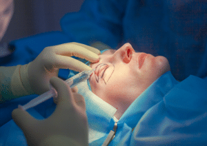
Local anaesthetic eye drops or sub-conjunctival anesthesia which is a local anesthetic injection into the eye will be used for this procedure before the cleaning of the wound and suturing is done.
Informed Consent:
For reconstructive surgery, the ophthalmologist will demonstrate the surgical repair process, possible medical risks and consequences, as well as post-operative care.
Patient Positioning:
The patient will likely be positioned in supine position.
Eyelid laceration surgery
Step 1: Preoperative evaluation:
Cleaning: Clean the wound with saline wash to clean out debris or wounds before proceeding to procedures.
Step 2: Canalicular Repair:
Identify both ends of the severed canaliculus. This requires the use of surgical loupes or an operating microscope to help visualize a surgical site better.
Use a monocanalicular or bicanalicular silicone stent such as Crawford or Mini Monoka stent at the cut ends for the lacerated canaliculus. The stent should pass into the lacrimal sac as well as through the nasolacrimal duct to keep it open as the tissues heal.
For bicanalicular stents, both the upper and the lower canaliculi are stented; the ends of the stents are then secured.
Close the ends of canaliculus with 7-0 or 8-0 polyglactin sutures that can be removed from the surface of the skin after surgery.
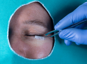
Step 3: Eyelid Repair:
Treat the tarsal plate, the muscle, and the skin, if involved, in layers in the repair process.
Non-absorbable sutures should be utilized, such as 6-0 silk or nylon.
Use absorbable sutures as 6-0 polyglactin and avoid using silk suture as it is not inert to tissue.
Use fine non-absorbable sutures for dermal approximation such as 6-0 or 7-0 nylon suture.
Pay attention to the eyelid margin as to avoid having a ‘notching’ or reposition.
Step 4: Postoperative Care:
Postoperative care after surgery
Antibiotics: It is recommended to treat the wound with topical antibiotic cream for the first day or two and administer oral antibiotics.
Anti-inflammatory Medication: It may be advisable to give medications such as Nonsteroidal anti-inflammatories, the goal is clearing of inflammation and tenderness.
Follow-Up: This should be accompanied with follow-up appointments frequently in an attempt to observe the healing progress and positioning of stents. The stent usually is left intact for 3-6 months.
Removal of Sutures: The skin sutures tend to be removed after between 5 to 7 days after the surgery has been performed.
Stent Removal: The silicone stent is taken out when the wound has healed sufficiently; normally this takes between 3 to 6 months but depends on the severity of injury and the surgeon who performs the procedure.
Infection: This is one of the most frequent manifestations of all types of wounds, including such injuries of the eyelids. In case contamination of the wound through a bite or a puncture occurs, the receiver is at a higher risk of contracting an infection. Signs of infection such as inflammation, pain, redness, and any discharge in form of pus.
Damage to the tear ducts: Tear ducts are a small tube which pump tears away from the eyes. If a wound reaches the tear producing tear ducts it means that the eye may either become too dry or begin to tear a lot.
Damage to the eyelid margin: The Eyelid margin is the edge of the eyelid and consists of the eyelashes; in this part of the eyelid that the eye-lashes are found. Injury to the eyelid margin results in wrong positioning of the eye lashes (trichiasis) that cause stickiness in the eye. It can also lead to issues with blinking, and how tears move through the eye.
Entropion: This refers to a turning inwards of the margin of the eyelid. This is more particularly hazardous as it can infect the eye, leading to irritation and corneal ulcers.
LEARNING & CME
View All
Advanced
Cardiovascular
Life Support

Basic Life
Support

Pediatric
Advanced Life
Support

Neonatal
Resuscitation
Program

Annual Stroke
Center
Continuing
Education

Opioid and Pain
Management

National
Institutes of
Health Stroke
Scale

Basics of
Electrocardiography