


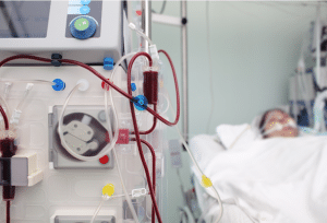
Extracorporeal Photopheresis is a more advanced therapy and is generally used for immunological related diseases. The procedure entailed the photochemical reaction of diseased cells and tumours by the reaction of a photosensitizing agent with blood cells and subsequent reinfusion of the agent under ultraviolet A (UVA) light into the patient.
Extracorporeal Photopheresis
Cutaneous T-Cell Lymphoma (CTCL) / Sezary Syndrome: ECP is FDA-approved for treatment of the advanced stages CTCL specially in Sezary syndrome which is the leukemic variant of CTCL. It is for the treatment of patients who fail to respond to other types of therapy.
Graft-Versus-Host Disease (GVHD): ECP is often employed for both acute and chronic GVHD particularly when patients may not respond to corticosteroids, other immunosuppressive treatments and in transplant immunization. It is also helpful in reducing steroid-dependent and symptom control in children.
Heart Transplant Rejection: ECP is done in combination with other immunosuppressive drugs in patients who have had recurrent or persistent allograft rejection.
Pemphigus Vulgaris: ECP has been used in the management of pemphigus vulgaris; this is a severe form of autoimmune blistering skin disease particularly when conventional therapies have failed to offer adequate solution.
Atopic Dermatitis: ECP can be therapeutic for patients with severe atopic dermatitis who do not benefit from other alternative treatments.
Photosensitivity disorders: Indications include the skin disorders like SLE and porphyria diseases that involve the skin among others as they increase the chances of adverse reactions from exposure to UV light.
Severe cardiac or pulmonary conditions: High spontaneous respiratory rates or minute ventilatory rates due to respiratory diseases could contribute to oxygen desaturation during the procedure.
Significant anemia or thrombocytopenia: When it comes to hemoglobin and platelet levels in patients, low levels may increase the risk of developing complications from ECP and the patient’s condition needs to be brought under control before ECP.
Apheresis Machine
Photoactivation Device
Photosensitizing Agent
Blood Collection and Infusion Sets
Support Equipment (centrifuge and incubators)
Comprehensive Review: First evaluate the patient’s medical background whether there have been any prior treatments, medications the patient is taking, and any other medical conditions.
Physical Exam: Assess the patient’s physical features to determine the presence of any potential risks that may make proceeding difficult or impossible.
Vein Evaluation: Assess whether a patient has access ports in the peripheral veins. Then the third option is the use of the central venous catheter.
Regular Medications: Note the regular medicines suggested for the patient and, if necessary, make changes to them. Some drugs may need to be stopped to avoid interaction with the drugs used in the procedure.
Photosensitizing Agents: It is advisable to refrain from medications that have the potential to magnify the effect of light as ECP employs UV radiation.
Step 1: Patient Preparation
Patient Assessment: Information is gathered from the conditions of the patient and his or her medical history. After that physical examination is done.
Consent: After that the procedure is explained to the patient allowing him/her to give informed consent.
Step 2: Venous Access
Venipuncture: An appropriate vein is identified, and the drug is administered through intravenous catheterization. If this cannot be achieved, a central venous catheter may be used, especially for patients with poor peripheral venous access.
Anticoagulation: Anticoagulants (e.g. heparin) are given to ensure that the arteries do not clot during the process.
Step 3: Blood Collection
Leukapheresis: Apheresis is a method in which leukocytes are obtained by first extracting blood from a patient and passing it through a machine that separates out the leukocytes from the rest of the blood.
Leukocyte Concentration: In the leukapheresis process leukocyte rich fraction is extracted from the blood while the rest of the blood components (RBC, plasma) are returned back to the patient.
Step 4: Photoactivation
Sensitization: 8-MOP is used as a photosensitizing agent for the collected leukocytes, and the mixture is exposed to light.
UV-A Irradiation: Leukocytes are exposed to ultraviolet A (UV-A) light after they are sensitized. This step activates the 8-MOP in the skin lesions to cross-link the DNA strands of the leukocytes leading to apoptosis.
Step 5: Reinfusion
Return of Treated Cells: Leukocytes that go through photoactivation are then returned to the patient’s system.
Step 6: Post-Treatment Monitoring
Observation: The concern is on whether there are adverse reactions to the procedures in the immediate.
Follow-Up: Follow-up involves visiting the patient and ensuring they are following the treatment well, and they are also being taken care of for any side effects that may arise after treatment.
Laboratory tests
Complete Blood Count (CBC): Check the blood levels in the patient since appropriate levels of platelets, red blood cells, and white blood cells are significant.
Liver and Kidney Function Tests: It is also advisable to monitor whether the liver and kidneys are doing their work properly; these organs are responsible for eliminating wastes.
Viral Serologies: Search for any possible infectious diseases such as hepatitis and HIV that can affect management.
Thrombocytopenia: Conversely, a decrease in platelet counts may contribute to the risk of bleeding, but this happens only rarely.
Skin Reactions: Exposure to UVA light and the photosensitizing agent can result in skin reactions.
Photosensitizing Agent Sensitivity: Allergic reactions to the photosensitizing agent 8-methoxypsoralen are not particularly common.
Thromboembolic events: Patients can also have clot formation at the venous puncture site.
Vascular Damage: Its use can damage veins and lead to problems such as stenosis or occlusion.
LEARNING & CME
View All
Advanced
Cardiovascular
Life Support

Basic Life
Support

Pediatric
Advanced Life
Support

Neonatal
Resuscitation
Program

Annual Stroke
Center
Continuing
Education

Opioid and Pain
Management

National
Institutes of
Health Stroke
Scale

Basics of
Electrocardiography