


End-tidal capnography is a medical monitoring technique that measures the concentration of carbon dioxide (CO2) at the end of an exhaled breath, commonly referred to as end-tidal carbon dioxide (ETCO2).
This monitoring method provides valuable information about a patient’s respiratory status and is widely used in various medical settings, including emergency medicine, anesthesia, critical care, and respiratory therapy.
During the normal breathing cycle, the body exchanges oxygen (O2) for carbon dioxide (CO2) in the lungs. Oxygen is inhaled, and carbon dioxide, a byproduct of metabolism, is expelled during exhalation.
Capnography involves the measurement of CO2 levels in the exhaled breath using a capnograph. The capnograph produces a waveform known as the capnogram, which represents the concentration of CO2 over time.
The capnogram typically consists of three phases:
Baseline (Phase I): Represents the beginning of exhalation, where little to no CO2 is present.
Exhalation (Phase II): Reflects the rapid rise in CO2 concentration as most of the exhaled air contains CO2.
Inhalation (Phase III): Shows a gradual decrease in CO2 concentration as fresh, CO2-poor air is inhaled.
- Capnograph Monitor
- CO2 Sampling Line
- CO2 Sensor
- Airway Adapter
- Informed Consent:
Before the procedure, assess the patient’s baseline respiratory status and overall health. Identify any pre-existing conditions that may affect ventilation.
- Patient Positioning:
Properly position the patient for the procedure and secure the capnography equipment. Ensure that the sampling line is appropriately connected to the patient and the monitor.
Step 1: Equipment Setup
Ensure that the capnography equipment is in good working condition.
Connect the CO2 sampling line to the patient.
Place the nasal cannula, endotracheal tube, or other airway device depending on the patient and procedure.
Step 2: Connection
Attach the CO2 sampling line to the airway adapter or the patient interface.
Step 3: Initialization
Turn on the capnograph monitor and allow it to initialize. This may involve a brief warm-up period.
Step 4: Baseline Assessment
Observe the initial baseline or Phase I of the capnogram, which represents the absence or low levels of CO2 at the beginning of exhalation.
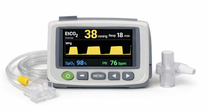
End-Tidal Capnography equipment
Capnography readings may be influenced by factors that can lead to false readings, such as the presence of ambient air, water, or other contaminants in the sampling line. This can result in inaccurate measurements of end-tidal CO2.
Malfunctioning capnography equipment, including the monitor, sampling lines, or CO2 sensors, can lead to unreliable or erroneous readings.
Improper placement of the CO2 sampling line can lead to inaccurate readings. In cases of severe airway obstruction or bronchospasm, the exhalation of gases may be impeded, potentially leading to a decrease in end-tidal CO2 levels.
During cardiac arrest or situations with low cardiac output, there may be a delay in the appearance of end-tidal CO2 in the exhaled breath, as blood circulation and CO2 delivery to the lungs are compromised.
LEARNING & CME
View All
Advanced
Cardiovascular
Life Support

Basic Life
Support

Pediatric
Advanced Life
Support

Neonatal
Resuscitation
Program

Annual Stroke
Center
Continuing
Education

Opioid and Pain
Management

National
Institutes of
Health Stroke
Scale

Basics of
Electrocardiography