


Electrodiagnosis is a field which makes use of Electrophysiology in the study of human neurophysiology with the aid of electricity. Neurodiagnostics(NDS), electromyography(EMG) and evoked potentials(EPs) are some of the fundamental principles that are used in electrodiagnosis.
Electrodiagnostic testing is a critical tool in evaluating peripheral nerve or muscle injury, muscle diseases, or the localization of the problem prior to developing a treatment strategy. While electrodiagnostic tests are functional, they fundamentally differ from structures like the MRI that provides an image of the body’s anatomy. For instance, a patient may experience severe pain at the back while the MRI scan may be normal; on the other hand, individuals may have disk protrusions but have no complaints. Thus, electrodiagnostic studies can be helpful in cases where other diagnostic modalities seem insufficient. There are physiatrists with specialization and training in physical medicine and rehabilitation, neurologists, anesthesiologists among others who are qualified to perform electrodiagnostic tests.
Electromyography (EMG) Machine: Captures electrical signals in muscles. Includes amplifiers, filters, and recording instruments to monitor and record signals picked by the needle electrodes that are inserted. Usually employed to identify muscle disorders and distinguish between neuropathies and myopathies.
Nerve Conduction Study (NCS) Equipment: Stimulates a nerve and records the velocity and amplitude of the signals sent across it. Uses surface electrodes mainly for stimulation and recording which is applied on the skin. Can evaluate the duration of nerve injury, conduction loss or demyelination.
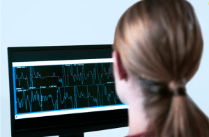
Evoked Potentials (EP) System: Captures EEG that shows the brain’s electrical activity in response to stimuli like visual/auditory/sensory. Applied in diagnosing diseases of the central nervous system including multiple sclerosis.
Patient preparation
Patients should be informed about the procedure and mild discomfort maybe felt during electrodiagnostic testing. They should be encouraged to wear free clothing so that they don’t have to struggle to get access to the sites for tests and should not apply any lotion or creams on their skin the day they are due for testing. Jewellery and metallic should be removed. It is also important for the patient to let the physician know if they have a pacemaker, any implantable devices or if they are on any medication especially anticoagulant. The skin should be free and clear of excessive moisture.
Patient position
Patients undergoing electrodiagnostic testing are usually in a comfortable sitting or lying position on the examination table or chair. In electromyography (EMG), the patient can be told to lie down or sit depending on the muscle that is being tested. Nerve conduction studies (NCS) require that the limb being examined be relaxed and immobilized, thus there is no muscle contraction. The position should make the patient comfortable and must be able to keep still for the entire length of the process to get a correct reading.
Electrodiagnosis
Step 1-Preparation: Security of a patient as well as their understanding of the process must be guaranteed. Wipe the skin at the electrode sites with alcohol swab to reduce the amount of oil at the electrode skin interface to enhance conductivity.
Step 2-Electrode Placement:
Surface Electrodes: In nerve conduction studies (NCS), adhesive surface electrodes are fixed on the skin over the nerves’ course.
Needle Electrodes: In case of electromyography (EMG), fine needle electrodes are then inserted into muscles to record electrical signals.
Step 3-Nerve Conduction Studies (NCS): Administer a low amplitude electrical current via surface electrodes. Evaluate the signal that is generated as a result following the propagation of the signal through the nerve and calculate the conduction velocity and amplitude, among other factors.
Step 4-Electromyography (EMG): Place needle electrodes into the muscle to monitor electrical activity both at rest and contraction. Superimposed on this background, assess SA, IA, and MUAPs to determine extant muscle function.
Step 5-Evoked Potentials: When assessing sensory tracts, use proper modality (visual, auditory, or somatosensory) stimulus and record electrical activity from electrodes positioned on scalp or other locations.
Step 6-Data Recording and Analysis: Record the signals using a computer and process them as displayed waveforms for interpretation purposes. Relative to reference values, one can note abnormalities. Step 7-Post-Procedure Care: You retire electrodes and clean the skin. Inform the patient on the instructions after the test and on the follow up depending on the results.
Complications
Discomfort or Pain: Pain sensations during needle insertions for electromyography (EMG), or when stimulating the nerve during nerve conduction studies (NCS), is moderate at worst.
Bruising or Hematoma: Percutaneous introduction of the needle can cause superficial haematomas and / or localized bleeding in patients with coagulation disorders or on oral anti-coagulant therapy.
Nerve Injury: However, there is potential harm to the nerve being tested; this is rare but more so with incorrect procedure or structural modification.
Infection: The chances of an infection at the area of needle insertion is relatively small, however, this should be compounded by poor aseptic techniques.
Allergic Reactions: There may be some complications if patients are allergic to substances like electrode gel or any local anesthetic if used.
LEARNING & CME
View All
Advanced
Cardiovascular
Life Support

Basic Life
Support

Pediatric
Advanced Life
Support

Neonatal
Resuscitation
Program

Annual Stroke
Center
Continuing
Education

Opioid and Pain
Management

National
Institutes of
Health Stroke
Scale

Basics of
Electrocardiography