


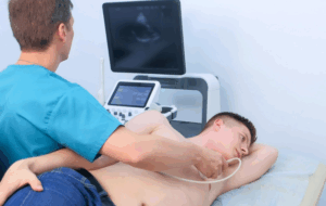
Stress echo or more specifically Dobutamine Stress Echocardiography (DSE) is a specific non-invasive test used for the assessment of response of heart muscles under stress or exertion. This test is mainly done on the patients with chest pain or CAD symptoms or any abnormalities that is related to the heart. Often, physical exercise might not be possible for some of the patients (because of limitation in mobility, in lungs or other conditions); thus, dobutamine, a medicine that has same effect on the heart as exercise, is given to set up the heart rate and echocardiography, which is an ultrasound scan of the heart to assess the function of the heart is carried out.
Dobutamine stress echocardiography
Diagnosis of Coronary Artery Disease (CAD): For identifying patients with ischemia or determining the extent of known CAD in coronary arteries.
Preoperative Risk Assessment: As a measure of cardiac risk in patients with known or suspected cardiovascular disease, prior to non-cardiac surgery.
Assessment of Myocardial Viability: Heart muscle viability testing in patients who have previously had a myocardial infarction or would develop heart failure and whether revascularization (as in surgery or stenting) is of use.
Evaluation of Heart Function: DSE for Left ventricular function assessment in patients with heart failure or cardiomyopathy.
Assessment of Valve Function: To evaluate valvular heart disease, especially aortic stenosis, and how the heart responds to stress.
Unstable Angina
For patients having unstable angina, the action of dobutamine is associated with increased myocardial oxygen demand and is consequently likely to precipitate another attack.
Severe Aortic Stenosis
Patients with severe aortic stenosis may develop sudden cardiac death during stress since their heart fails to deliver blood through the narrowed aortic valve at increased demand.
Uncontrolled Hypertension
Dobutamine stress causes hypertensive crisis and stroke when blood pressure is higher than 180/110 mm Hg.
Obstructive hypertrophic cardiomyopathy
This is moderately hereditary and caused by variation in the tropomyosin gene. The contractility enhanced with dobutamine decompensates these patients and increases the outflow obstruction leading to ventricular dysrhythmias or worsened symptoms.
Severe Arrhythmias
Dobutamine has adverse effects on patients with pre-existing arrhythmias; PVCs, ventricular tachycardia, and uncontrolled atrial fibrillation to life-threatening events.
Echocardiography Machine
Dobutamine Infusion Pump
Probes (5 Hz,3.5 Hz)
Contrast agitator machine
Echo beds
Monitoring Equipment:
Resuscitation Equipment (for emergencies):
Software for Image Analysis:
Medication review: The physician should also consider changing some of those drugs, especially beta-blockers, as they interact with dobutamine.
Allergies: Confirm to the healthcare team the existence of any drug allergies especially to dobutamine or any medication to be used during test.
Clothing:
Avoid wearing tight clothes, wear comfortable clothing. Patients may be required to wear a hospital gown.
Informed Consent:
Sign consent forms before the procedure. The healthcare team will discuss with the patient the dangers of the procedure and its advantages as well as the other options in case the procedure is considered.
Patient Position:
The patient is usually placed in a left lateral decubitus position, or on their left side, or in a supine, that is, on their back.
Step 1-Preparation:
Patient Assessment: Obtain a detailed past medical, medication and social history, current symptoms and physical signs. Contraindications include severe aortic stenosis, recent myocardial infarction or severe arrhythmias.
Informed Consent: The patient should be informed about the procedures and the possible risks involved in the procedure.
Baseline Echocardiography: The first test to perform is an echocardiography to determine the patient’s baseline cardiac function and anatomy.
Equipment Setup: Perform an echocardiographic examination using ultrasound machine that has capability to produce.
Doppler signals to evaluate the motion of the myocardial and blood. Order to set an I.V. line for dobutamine administration to start.
Step 2-Dobutamine Administration:
Dosage and Infusion Protocol: Dobutamine is usually given at a low starting rate (for example 5 µg/kg/min), however this can be increase to optimum rate of 40 µg/kg/min every 3 minutes depending on the patient condition and heart rate.
Monitoring: Continuous monitoring the pulse, & ECG need to be inspected constantly throughout the process. Monitor for side effects such as angina or any change in rhythm of heart.
Step 3-Echocardiographic Imaging Image Acquisition: Start echocardiographic investigation both at baseline and during dobutamine stress. In all patients, standard views must be acquired to assess wall motion and function, such as the parasternal long axis, parasternal short axis, apical four-chamber view, and apical two-chamber views.
Step 4-Assessment of Myocardial Perfusion: It’s recommended to use Doppler imaging to evaluate blood flows and wall movements irregularities.
Step 5-Termination of the Test: The test may be stopped if the patient’s symptoms worsen, patient’s predicted maximal heart rate is achieved, or if values on the ECG change significantly, for example ST segment depression.
Dobutamine infusion is stopped, and the patient must be closely observed until the heart rate and blood pressure get stabilized.
Dobutamine infusion is discontinued, and the patient should be monitored until their heart rate and blood pressure return to baseline levels.
Step 6-Post-Procedure Care:
Possible side effects of dobutamine should be looked for and the patient should be stable out before discharge.
Discharge instruction covering signs and symptoms to report in the next following days after the procedure.
Step 7-Interpretation of Results:
Wall Motion Abnormalities: Ultrasonography images of the heart to look for regions of the heart where wall thickening, or motion is decreased or abnormal motion-this would signify ischemia.
Functional Capacity: Supervise function of the left ventricular before and after giving Dobutamine thus ascertain whether normal function is regained.
Diagnosis: They see if there is a chance of developing coronary artery disease because of presence of wall motion abnormalities resulting from stress.
Arrhythmias: The most common complication, including premature ventricular contractions (PVCs), atrial fibrillation, or other supraventricular tachyarrhythmias. These can occur due to increased heart rate and myocardial oxygen demand.
Hypotension: It can happen because dobutamine can cause vasodilation, in patients who have weak blood vessels or if they are dehydrated.
Myocardial Infarction: Occasionally, the myocardial perfusion imaging stress test can provoke a myocardial infarction in patients who apparently have severe coronary artery disease if the patient experiences major ischemia during the test.
Cardiac Arrest: However, such complications are very rare but can happen in patients with cardiac disease or those with severe blockage of the arteries.
Pulmonary edema: In some patients, especially in those who developed heart failure in the past, dobutamine may lead to the worsening of pulmonary congestion.
LEARNING & CME
View All
Advanced
Cardiovascular
Life Support

Basic Life
Support

Pediatric
Advanced Life
Support

Neonatal
Resuscitation
Program

Annual Stroke
Center
Continuing
Education

Opioid and Pain
Management

National
Institutes of
Health Stroke
Scale

Basics of
Electrocardiography