


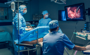
Cystoscopy is a surgery or an examination that is done through the urethral using an endoscopic tube. This can be accomplished with a rigid or a flexible cystoscope and is for both diagnostic and treatment functions.
The internal body visualization can be dated back from as early as 1807 when Philipp Bozzini, a German military surgeon, invented the Lichtleiter, which was an early model of endoscope to seek for bullets in the patients. The current cystourethroscopes feature optimal light source in the form of cold light illumination, enhanced lens resolution, video endoscopy coupled with flexible instruments and lastly virtual endoscopy.
In adults, the bladder lies in the anterior abdominal wall in the pelvic region and is enclosed within the extraperitoneal fat and connective tissue. This is located at the anterior aspect of the lower abdomen, just behind the pubic symphysis with the retropubic space or space of Retzius in-between. Typically, the upper part of the bladder has peritoneal lining while the lower most part is fixed by pelvic fascia and ligaments. From below it is bounded by external urethral sphincter and perineal membrane, and laterally by two muscles of obturator internus.
Cystoscopes are of two types flexible and rigid, and their size is based on French (Fr) gauge. In rigid cystoscopes, Hopkins rod-lens system provides better vision than the standard images; on the contrary, flexible cystoscopes provide more comfortable vision to the patient and easier in movements irrespective of their type and nature of procedures taken place in cystoscopy. Rigid scopes offer better image quality and larger channel for instrument compared to flexible scopes, but flexible scopes are smaller and easier in specific positions.
Rigid Cystourethroscopy: Special rigid scopes used by companies Karl Storz and Olympus have lenses with the range from 0 to 120 degrees and are complemented by bridges and sheaths. Small sheaths are used for diagnosis to minimize injury to a vessel while larger sheaths are employed for therapy.
Flexible Cystourethroscopy: There are a variety of flexible cystoscopes with parameters referring to the diameter which can range from 16-17 Fr; the models differ in the controllability of the tip, optics, and light source. Modern digital models have higher resolution than older analog ones and much better light resolution than fibreoptic models but possess worse depth of field.
Irrigants: Water is in most cases normal saline, and sterile water can also be used for irrigation. Monopolar electrocautery must use a non-ionic fluid to prevent harm to tissues and bipolar electrocautery must use isotonic fluid and nothing else. In cytology sample collection, sterile water is an important component.
Patient preparation
A cystoscopy is a medical procedure, and it is mandatory to obtain a clear consent from the cystoscopic patient. Prior to starting with an antiseptic wash, a urinalysis and urine culture are usually done. Antibiotics are usually not required before cytoscopic surgery based on AUA guidelines; however, patient-related risks should be considered before performing the procedure.
These risk factors include:
Advanced age Pathological conditions of the urinary System
Chronic corticosteroid use
Some of the colonized material that could be present are endogenous and can be drawn from the GM or from outside the GM.
Distant coexisting infection
Immunodeficiency
Poor nutritional status
Persistent infections
Smoking history
With therapeutic procedures, a short term (24 hours or less) prophylaxis with a fluoroquinolone or trimethoprim-sulfamethoxazole should be given. Other choices are an aminoglycoside with or without ampicillin, a first- or second-generation cephalosporin, or amoxicillin and clavulanic acid.
Patient position
In cystoscopy, the position of the patient depends on the type of procedure to be conducted and the type of cystoscope to be used. Lithotomy position is used in rigid cystoscopy where patient lays supine with legs flexed at the hips and supported by stirrups with the patient’s buttock off the edge of the table while the thighs are raised with a turn to ensure easy visualization of the urethra and bladder. At times, frog leg position can also be used in which the ankles of the patient are flexed, and thighs are apart to allow access that increases comfort. For flexible cystoscopy the patient lies flat on his back in supine position and the cystoscopy is much easier with a flexible instrument. Ideally positioning require a lot of attention to ensure that both the patient and the surgical procedure are well positioned.
Step 1-Preparation: Examine the external genitalia for evidence of any lesion or anatomical distortion prior the start of the procedure.
Step 2-Positioning: Specifically, for women, a sheath obturator should be used while performing rigid cystourethroscopy. Size the scope while positioning it further ahead in the direction of bladder. For men, be sure that the penis is elongated as much as possible to align the urethra in a straight line.
Step 3-Handling the Penis (Men): Pul the penis with the left-hand which forms the working hand (five finger grip for rigid cystoscopy). For flexible cystoscopy gently grasp the penis between the third and fourth fingers of the non- dominant hand, leaving the thumb and forefinger to manipulate the scope. Rotate the penis 45 to 90° to the abdominal wall as the scope is being passed through the anterior urethra.
Step 4-Advancing the Scope: Having crossed through the membranous urethra turn the scope in an anteromedial direction to access the bladder:
In the case of subjects with flexible scopes use active upward flexion only.
For rigid scopes, lower the distal end of the scope toward the operative table.
Step 5-Evaluation of the Lower Urinary Tract: Respectively scanning will systematically evaluate the lower urinary tract while maintaining that the maximal irrigation is running. In the penile and bulbar urethra look for stricture or any other anomaly. Comfort the patient when the scope passes through the membranous urethra as it continues the journey.
Step 6-Identifying Key Structures: In the prostatic urethra state the verumontanum and the utricle. Examining the size of the prostatic lobe, the length of the prostatic urethra as well as the presence of a median lobe of bladder neck dorsum.
Step 7-Bladder Inspection: After initiating inspection of the bladder, use the 30-degree scope first to have a view on as many parts of the bladder as possible. Assess the bladder floor and trigone to determine the position and the number of ureteral orifices as well as searching for blood efflux.
Cystoscopy
Step 8-Comprehensive Bladder Examination: Examine the bladder for presence of stones or any trabeculations and diverticula, and any erythematous patches or any individual papillary/sessile lesion. Tilt the cystoscope so that the lateral walls are displayed on the screens, the position of camera should remain the same. In rigid endoscopy, use a 70 or 120-degree lens while in flexible endoscopy perform retroflexion to view the dome and posterolateral walls.
Step 9-Final Steps: It is also important to have the bladder emptied before the removal of the scope.
Technical considerations
The antibiotic prophylaxis should be prescribed only to the patients most at risk of developing UTIs, hence simple cystourethroscopy may be followed by antibiotic prophylaxis only in cases if the patient has risk factors, such as age, urinary tract anomalies, poor nutrition, smoking, the use of corticosteroids, immunodeficiency, the presence of catheters, infections as well as hospitalization for the scope of the procedure. In these cases, a dose of fluoroquinolone or of trimethoprim-sulfamethoxazole; other choices include aminoglycosides, cephalosporins or amoxicillin-clavulanate. Those with negative urine culture and no risk factors do not require prophylaxis. Although these guidelines have been developed for patients with simple cystourethroscopy, all patients who undergo procedures that involve manipulation should be given antibiotic prophylaxis. The American Heart Association has also stressed that antimicrobials are no longer used as prophylactic as in the case of genitourinary procedures with a view to preventing infectious endocarditis.
Laboratory tests
Complete blood culture
Urinalysis
Urine culture
Ultrasound
CT scan
Complications
Possible risks of cystoscopy are normally minimal and may include UTI, hematuria, dysuria and possible injury to the bladder or the urethra. A known consequence is an iatrogenic urethral stricture brought about by the instrumentation employed during the surgery.
LEARNING & CME
View All
Advanced
Cardiovascular
Life Support

Basic Life
Support

Pediatric
Advanced Life
Support

Neonatal
Resuscitation
Program

Annual Stroke
Center
Continuing
Education

Opioid and Pain
Management

National
Institutes of
Health Stroke
Scale

Basics of
Electrocardiography