


Cardiac ultrasound is sometimes called echocardiography which is a therapy that involves using sound waves to generate images of the heart. As such, it is a non-invasive and safe procedure which evaluate the anatomy and functioning of the heart.
It is based on the principles of ultrasound technology, it uses high-frequency sound waves to create images of internal organs.
Echocardiography involves use of high-frequency sound waves (ultrasound) produced by a transducer placed on the skin of the chest. These sound waves then move through the body then if they reach a structure like the heart, some are reflected to the transducer. These returning sound waves are then converted into images by using real time view of the heart’s anatomy and functionality.
Equipment
Ultrasound Machine
Ultrasound Transducer
Electrocardiogram System
Gel
Patient Monitoring Equipment
Patient Preparation
Informed Consent:
Explain the risks and benefits of the procedure to the patient and obtain informed consent.
Patient Positioning:
For a standard echocardiogram, position the patient comfortably on an examination table resting on their left side while in the process of a transesophageal echocardiogram (TEE), the patient should sit with semi upright position.
Cardiac ultrasound
There are two primary types of cardiac ultrasound: transthoracic echocardiography (TTE) and transesophageal echocardiography (TEE).
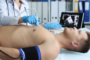
Step 1: Application of Gel:
Preparing the surface of the patient’s chest, a water base gel is used. As a result, the transmission of ultrasound waves is enhanced, and clear visualization is achieved.
Step 2: Placement of Transducer:
Ultrasound transducer emits and receives ultrasound waves that are positioned in different locations on chest to capture different chambers of the heart. Parasternal, apical and intercostal oblique, and subcostal views are employed to capture optimal images of the heart.
Step 3: Image capturing
Take enough images to evaluate chambers, valves, and determine the function of the heart.
Step 4: Probe Manipulation
In some cases, the patient must switch positions or, breathe in a certain way to get optimal image quality and to evaluate different aspects of cardiac performance.
Various views allow for a detailed examination of different aspects of the heart as follows:
Parasternal long-axis view: It is a side view of the heart, showing the left ventricle, aorta, and mitral valve.
Apical Four-Chamber View: This takes pictures of all four chambers of the heart (left atrium, left ventricle, right atrium, and right ventricle).
Apical Two-Chamber View: The left atrium and ventricle of the view that is shown provide a longitudinal view of the left side of heart.
It supports the four-chamber view in its entirety to give a more complete assessment.
Mid-esophageal four-chamber view: It is essentially the transthoracic apical four-chamber view but obtained through the esophagus.
Trans gastric Short-Axis View: Obtained by angling the TEE probe into the stomach by providing a short-axis view of the left ventricle.
4.Complications
Complication arising from sedation includes respiratory suppression, allergy on the sedation agents used and effects arising from sedation on specific respiratory disease.
Insertion of the TEE probe may cause minor damage to the esophageal lining.
Formation of a perforation, a very rare but serious complication, may also occur.
The gag reflex is elicited in TEE, and if previous measures are not applied, aspiration can occur.
There are possibilities of these changes being more sensitive to cardiovascular illnesses patients. The TEE probe is placed in the mouth, it rarely causes dental damage, but if a bite block is not used, or there is pathologic change in the teeth.
LEARNING & CME
View All
Advanced
Cardiovascular
Life Support

Basic Life
Support

Pediatric
Advanced Life
Support

Neonatal
Resuscitation
Program

Annual Stroke
Center
Continuing
Education

Opioid and Pain
Management

National
Institutes of
Health Stroke
Scale

Basics of
Electrocardiography