


Lymphoscintigraphy or sentinel lymph node mapping is an imaging method which is used to detect the lymph drainage basin, the number of sentinel node, distinguish sentinel node from subsequent node, locate the sentinel node in a random location, and identify sentinel node on skin for biopsy. Lymphoscintigraphy is suggested to verify nonpalpable or palpable invasive breast cancer. It needs primary tumor removal and dissection of axillary node.
Sentinel node mapping is fast emerging method as an alternate staging approach for the axilla in the treatment of early breast cancer. It involves the injection of a radioactive material into the body. It travels into the lymphatic system. A specialized camera detects the material and records photographs of its passage. Lymphoscintigraphy is used to identify the location of the sentinel lymph node (SLN) which is first lymph node to which cancer cells are most likely to disseminate. This can guide in the biopsy. The test can also detect obstructions in lymphatic drainage system.
Imaging system and gamma camera
Gamma detecting probe
Radiopharmaceuticals substance: Technetium Tc-99m: antimony trisulfide with particle size of 0.015 to 0.3 µm, nanocolloid with particle size of 0.05 to 0.8 µm, or sulfur colloid with particle size of 0.22 µm
Syringe with needle of proper gauge (most usually 25 G insulin syringe)
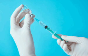
Injecting syringe
Syringe shied
Alcohol swaps
Monitoring equipment to check the vital signs of patient
Patient preparation
No anesthesia is needs.
Patient must drink plenty of water to stay hydrated during the procedure and increase the accuracy of procedure.
Patient must inform to the healthcare providers if they are taking any anticoagulants or blood thinner medications before the procedure.
Patient position
Radiopharmaceutical injection positions:
Patient must be positioned in the supine position on the imaging table with exposed breast area. Administer the injection near the tumor site subdermally or intradermally.
Position during imaging methods: Gamma camera is positioned above or around the patient for the imaging. The patient may have to change the position to get the images from different angles.
Intradermal injection method:
A 0.2 mL injection of 10 to 15 MBq (0.3 to 0.4 mCi) of Tc-99m substance is made up before the 24 hours of surgery.
A 0.5 mCi dosage of Tc-99m tilmanoceptis given before 15 minutes of intraoperative lymphatic mapping.
A 25-gauge needle is used with 0.2 mL air bubble to draw the syringe. It reduces the risk of leakage. The injection is given on the skin which has tumor and needle is inserted at the proper angle in the skin. If the injection method is proper, it is confirmed by the appearance of skin bleb at injection area.
Dry cotton is used on the injection site and it is sealed with adhesive bandage. Patient has given instruction to give massages at the injection site for 1 to2 minutes with dry cotton wool.
Peritumoral injection method:
The peritumoral injection method includes the injection in the breast parenchyma near the lymph.
About 4 injections are given near the tumor. The volume of the injectate is increases with 1 to 2 mL at each injection area of a total volume of the 4 to 8 mL.
The injections are given inferior, superior, medial or lateral to bulk. Another technique is to inject in semicircle with axillary side of tumor.
Needle must be replaced with other one with ultrasound if the lump is not palpated.
The patient has given some instructions to massage for 1 to 2 minutes between the axilla and injection area.
Intratumoral injection technique
A 0.2 mL of radiopharmaceutical with 0.1 mL of air bubble is injected in the center of tumor. Needle must be replaced with other one with ultrasound if the mass is non-palpable.
The patient has given some instructions to massage for 1 to 2 minutes between the axilla and injection area.
Patent blue, isosulfan blue or methylene blue injections are utilized separately or along with others to identify the sentinel node. It is given before the 5 minutes of the surgery.
It can be given peritumoral, subareolar, or subdermal. A 1.5 to 2 mL of volume is used for the subdermal injection. A large volume in the 2 aliquots total 5 mL is used for the peritumoral injection.
The injection area is massages as it contains radiopharmaceutical substance.
When a blue color appears, it leads to positive result.
Sentinel Node Imaging for Breast Surgeons:
It gives a visual help to fetch sentinel node. It gives radioguidance with the gamma probe for interdisciplinary operation. It assists o determine the area of sentinel node.
It counts the area after the removal of the full excision. It guides the surgeon to remove the non-blue nodes with the radioactive signature.
The gamma probe is protected with the sterile sheath. The sight is established in the angle which has eh short distance from the skin to sentinel node.
The node is contacted along its line of sight, with the probe providing periodic feedback to adjust the direction. If a blue color appears, it confirms the direction.
Once the node is seen, probe is applied to make sure the high count rate. If blue dye is utilized and same color is appeared, there is confines the probe data.
The probe is re-used on the surgical site to confirm the radioactive node is removed. IF all the nodes are removed, then the surgical bed activity will decrease to 10% of the active node.
Complications:
Injection site reactions like infection or inflammation
Allergic reactions
Exposure to radiation
False negative or false positive results
Lymphedema
Discoloration
Rare cardiac complication
Discomfort during imaging
LEARNING & CME
View All
Advanced
Cardiovascular
Life Support

Basic Life
Support

Pediatric
Advanced Life
Support

Neonatal
Resuscitation
Program

Annual Stroke
Center
Continuing
Education

Opioid and Pain
Management

National
Institutes of
Health Stroke
Scale

Basics of
Electrocardiography