


Bipedicle Transverse Rectus Abdominis Myocutaneous (TRAM) flap breast reconstruction is a procedure that is being done in most cases where the breast needs to be reconstructed after the mastectomy. Conversely, the tissue expander technique entails with using the tissue from the lower abdomen area where the doctor creates the breast mound. This term bipedicle means the flap is supported by two blood supplies (pedicles) that support the tissue flap.
- Surgical Instruments
- Microsurgical Equipment
- Microscope or Loupes
- Suture Material
- Drapes and Sterile Supplies
- Anesthesia Equipment
- Operating Room Setup
- Postoperative Care Supplies
Preoperative consultation: This is the important point; you should discuss your aims and expectations with your surgeon. They will evaluate if you are fit for the procedure by examining your current health, body type and availability of donor’s tissue type.
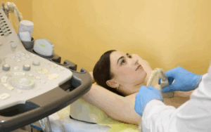
Preoperative examination of breast
Medications: In some cases, the doctor may need to temporarily discontinue certain medications or adjust before surgery. Medical providers recommend you consult the doctors regarding all prescribed drugs.
Patient position
The patient is typically positioned in a flexed supine position.
Step 1: Pre-operative planning and evaluation:
The consultant should perform various assessments to determine patient expectations, medical history, and the possibility of performing the procedure.
While, imaging scans such as mammogram and CT scan could be done to evaluate the chest and abdominal wall.
Step 2: Incision and flap harvest:
The surgery is generally conducted with the administering of the general anesthesia.
The slit-like cuts are made at the lower portion of the abdomen with one cut above and one below the navel.
The flap (panniculus) and the rectus muscle elevates together, puling the tissue taut along with the blood flow from the rectus muscle.
At its upper and lower ends, the rectus muscle will be transected. This leads to central portion subsisting within intact abdominal wall strips.
The vessels running on the flap (perforators) are precisely located and spared.
Step 3: Flap transfer and recipient site preparation:
The abdominal flap is transferred to chest wall.
The nipple and areola reconstruction may be completed at the same time as the reconstructive surgery. However, it can sometimes be done in a separate procedure.
Here, the donor site, drawn on the chest wall, is ready for the transfer of the flap.
Step 4: Microvascular anastomosis:
The blood vessels are microvascular on the flap from the base of the chest wall are also carefully anastomosed (connected) to the blood vessels of the internal mammary artery and vein.
Step 5: Flap shaping and closure
Sculpting and positioning the flap to achieve the desired breast shape and size, along with reshaping and repositioning the umbilicus.
It is possible to repair the areola and nipple in a different procedure.
Sutures will be used to close the abdominal and chest wall incisions.
Complete Blood Count (CBC): This could involve diagnosing anemia and infections among other. Where blood disorders are concerned, they make possible the identification as well as potential cause of the disease.
Basic Metabolic Panel (BMP): It is to assess the GFR (take show the kidneys function), then measure the electrolyte level (sodium, potassium, chloride), the blood sugar, and the liver functionality.
Flap Necrosis: Sometimes the tissue grown for the breast construction may be unnourished adequately and, as a result, some tissue cells may die (necrosis). The partial or entire breast (escalation) may be lost, thus it should be considered as the resultant part.
Seroma Formation: Seroma is a fluid or serum formation that can sometimes be seen in the surgical site. Though most of them end by the time, a seriously or chronically sized seroma may require drainage.
Infection: Infection is the risk of most of surgeries, hence it will also be a result of TRAM breast reconstruction surgery.
LEARNING & CME
View All
Advanced
Cardiovascular
Life Support

Basic Life
Support

Pediatric
Advanced Life
Support

Neonatal
Resuscitation
Program

Annual Stroke
Center
Continuing
Education

Opioid and Pain
Management

National
Institutes of
Health Stroke
Scale

Basics of
Electrocardiography